
Clinical Review Notice: Adapted from the Joint & Orthopedic Center at Myongji St. Mary's Hospital. This publication outlines knee joint diagnostic protocols, stratified treatment frameworks, and preventative guidelines.
💡 Quick Summary: At a Glance
- Multi-Modal Diagnosis: Specialized orthopedic evaluation combines X-rays, 3D CT scans, high-resolution MRI, and diagnostic arthroscopy to pinpoint precise soft tissue and cartilage damage.
- Stratified Treatment Tiers: Care progresses systematically from Tier 1 (conservative RICE care) and Tier 2 (NSAIDs, hinged bracing, hyaluronic acid injections) to Tier 3 (Arthroscopy, HTO, or Total Knee Arthroplasty).
- Lifelong Joint Protection: Preserving joint health requires weight control, leg muscle strengthening, dynamic warm-ups, and strictly avoiding high-stress floor postures.
🎯 Who This Guide Is For: This clinical guide is written for individuals evaluating diagnostic imaging, non-surgical treatment options, or surgical interventions for knee pain who want a structured roadmap for joint care and recovery.
Looking for Causes & Symptoms? If you have not yet identified the underlying cause of your knee pain, read 👉 Part 1: Common Knee Pain Causes & Symptoms Identification Guide.
1. Multi-Modal Diagnostic Protocols for Knee Joint Damage
Accurate knee joint diagnosis requires combining weight-bearing X-rays with soft-tissue MRI scans and specialized 3D imaging to pinpoint structural cartilage and ligament injuries.
Because clinical presentation varies significantly from patient to patient, specialists at Myongji St. Mary's Hospital utilize a multi-modal diagnostic process to ensure targeted, effective intervention:

- Clinical History & Physical Exam: Evaluating point tenderness, joint laxity, patellar alignment, range of motion, and performing specialized orthopedic stress tests.
- X-Ray Examination: Assessing bone alignment, joint space narrowing, structural deformities, and bone spur (osteophyte) formation.
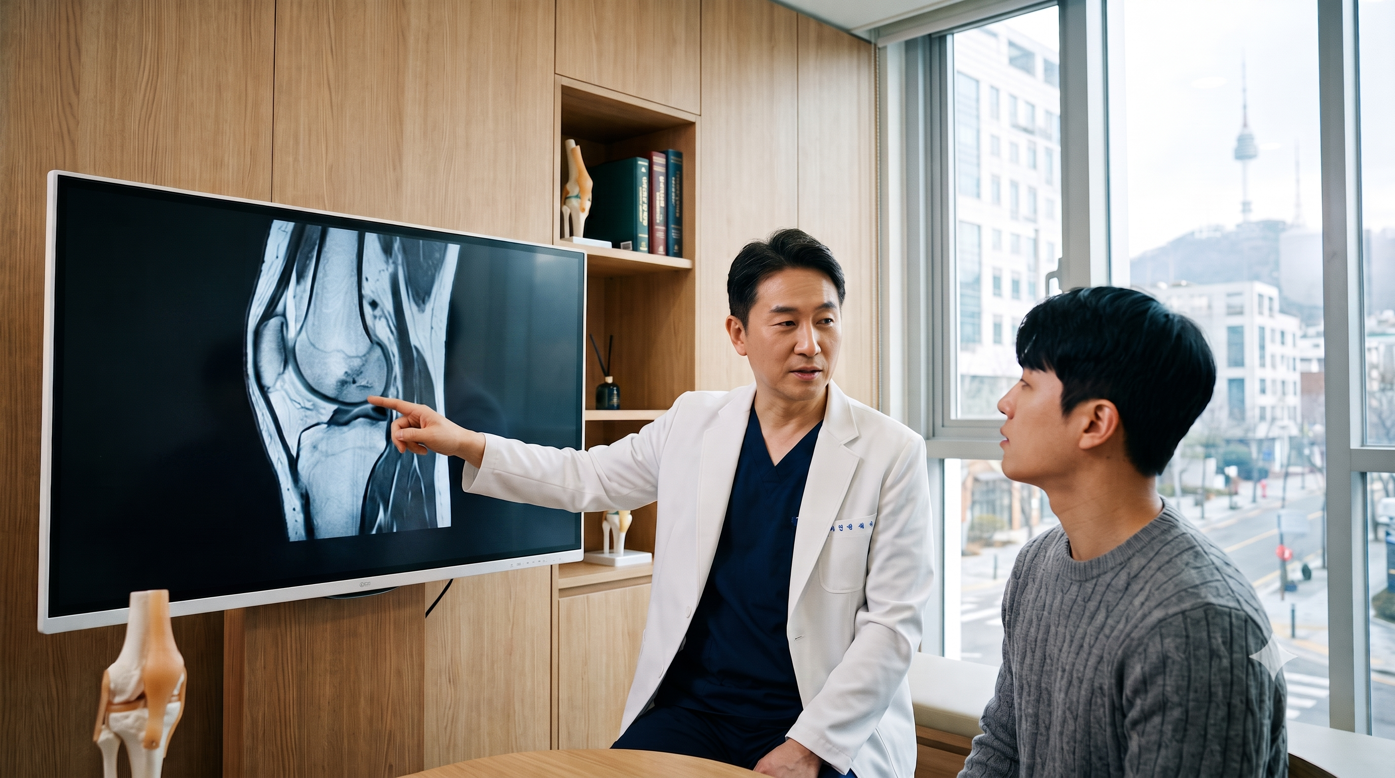
- Magnetic Resonance Imaging (MRI): The gold standard for confirming the exact extent of soft tissue disruption, including meniscal tears, cartilage wear, and ligament ruptures.
- Computed Tomography (CT) Scan: Providing detailed three-dimensional structural mapping for complex intra-articular bone trauma or alignment deformities.
- Radioisotope Bone Scan: Screening for subtle bone stress responses, metabolic changes, deep bone marrow edema, or occult infections.
- Diagnostic Arthroscopy: Inserting a miniature endoscopic camera directly inside the joint space. Used in complex or ambiguous cases where non-invasive imaging is inconclusive, allowing direct visual inspection and immediate treatment of internal structures.
2. Stratified Treatment Framework (Tiers 1 through 3)
Orthopedic care progresses systematically through three distinct tiers, starting with conservative self-care, advancing to non-surgical medical interventions, and reserving surgery for structural failure.
Treatments are assigned systematically based on symptom duration, functional impairment, and structural damage severity.
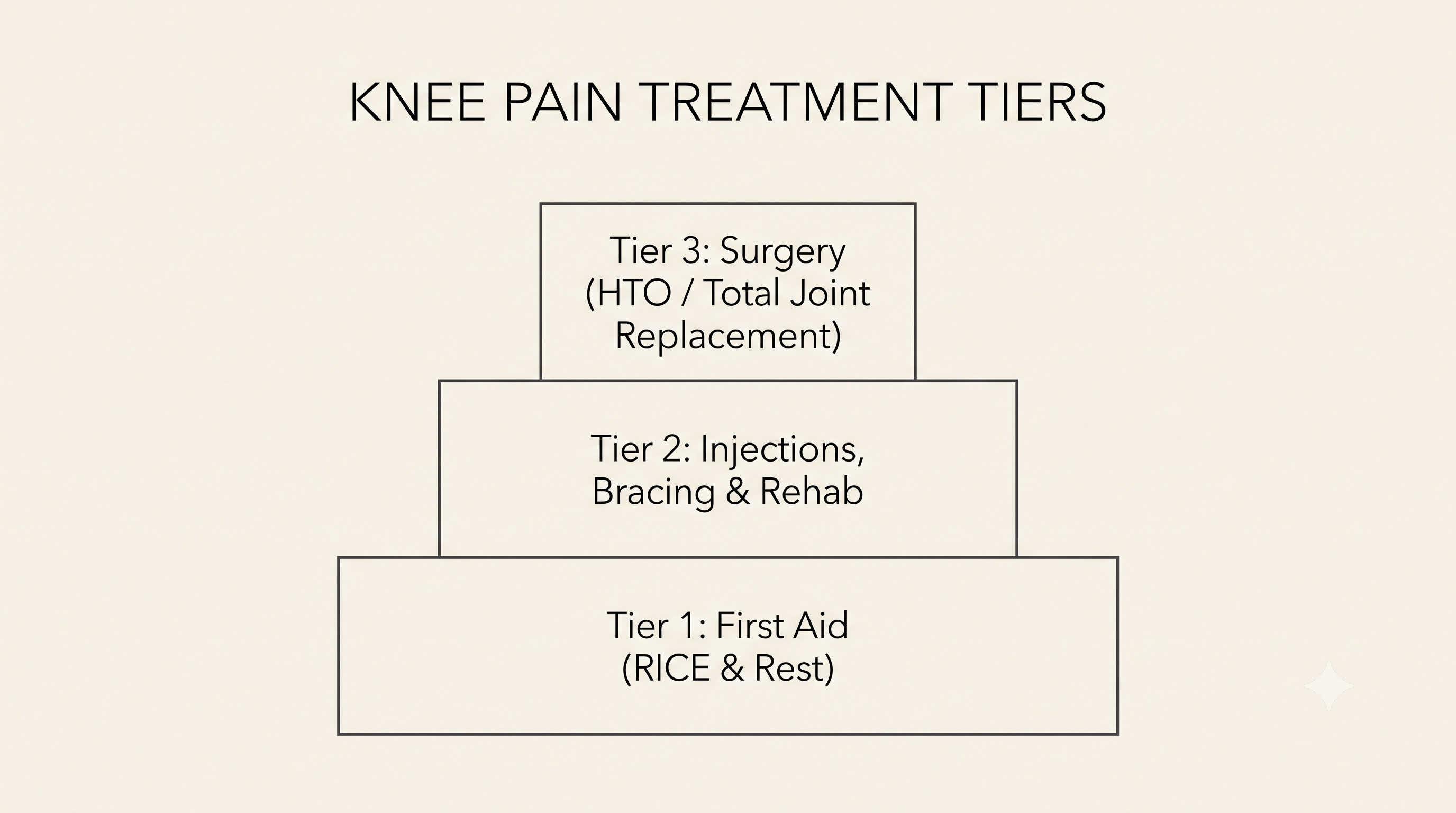
Figure 2: Stratified Treatment Framework—stepwise progression from conservative RICE protocols to advanced surgical interventions.
| Treatment Tier | Severity & Clinical Indication | Interventions & Specific Therapies |
|---|---|---|
| Tier 1: Conservative First Aid | Acute minor strains, overuse irritation, or initial mild pain flares. |
• RICE Protocol: Rest, Ice, Compression, and Elevation. • Activity Modification: Temporary load reduction to prevent micro-trauma progression. |
| Tier 2: Non-Surgical Medical Care | Moderate structural damage or persistent symptoms resisting self-care. |
• Joint Immobilization: Custom splints or hinged knee braces. • Pharmacotherapy: Oral anti-inflammatory medications (NSAIDs). • Targeted Injections: Intra-articular hyaluronic acid (viscosupplementation gel) or corticosteroids. • Physical Rehabilitation: Manual therapy and targeted quadriceps/hamstring strengthening. |
| Tier 3: Advanced Surgical Intervention | Severe structural disruption, failed conservative care, or end-stage joint arthritis. |
• Arthroscopic Surgery: Minimally invasive keyhole repair or debridement. • High Tibial Osteotomy (HTO): Realignment of the shinbone to relieve pressure on single-sided osteoarthritis. • Total Knee Arthroplasty (TKA): Full artificial joint replacement for severe, chronic pain impairing daily living. |
Tier 1: Conservative First Aid (Acute Strain Care)
For initial mild pain flares or acute minor strains, the immediate focus is reducing localized tissue inflammation through the RICE protocol (Rest, Ice, Compression, and Elevation) alongside temporary load reduction to prevent micro-trauma progression.
Tier 2: Non-Surgical Medical Care & Injections

Figure 3: Tier 2 Rehabilitation—targeted physical therapy focusing on leg muscle strengthening to absorb joint impact.
When moderate pain or structural irritation resists basic self-care, medical intervention stabilizes the joint and restores mobility:
- Joint Immobilization: Custom splints or hinged knee braces to control unnatural side-to-side motion.
- Targeted Injections: Intra-articular hyaluronic acid injections (viscosupplementation gel) to lubricate joint surfaces or short-term corticosteroids to quiet acute inflammatory flare-ups.
- Physical Rehabilitation: Specialized physical therapy focusing on quadriceps and hamstring muscle strengthening to absorb mechanical shock.
Tier 3: Advanced Surgical Intervention
When conservative therapies fail to provide relief, or when severe structural disruption exists, surgical intervention restores mechanical alignment and function:
- Arthroscopic Surgery: Minimally invasive keyhole procedure used to trim torn meniscal edges, clean out loose cartilage debris, or reconstruct torn ligaments.
- High Tibial Osteotomy (HTO): A joint-preserving surgical realignment of the upper shinbone. By correcting leg angle (e.g., bowleg alignment), weight is shifted away from the worn inner joint compartment onto healthy outer cartilage.
- Total Knee Arthroplasty (TKA): Full artificial joint replacement designed for end-stage osteoarthritis where joint cartilage is completely worn away, restoring pain-free walking and quality of life.
3. The 5-Point Knee Protection Protocol for Lifelong Mobility

Figure 4: Dynamic Joint Warm-Up—stimulating synovial fluid release to lubricate cartilage surfaces prior to physical activity.
Preserving joint cartilage and preventing progressive degradation requires proactive weight management, quadriceps strengthening, dynamic warm-ups, and eliminating compressive floor postures.
Preventing progressive joint degradation and protecting your knees after receiving medical treatment is essential for maintaining lifelong mobility.
- Maintain a Healthy Body Weight: Reducing body mass significantly decreases the exponential mechanical load placed across joint cartilage during daily walking and stair climbing.
- Perform Dynamic Warm-Ups: Engage in active movements (leg swings, light squats) before workouts. Warm-up activity stimulates the release of synovial fluid—your knee’s natural joint lubricant—allowing cartilage surfaces to glide smoothly without painful friction.
- Control Workout Intensity: Increase exercise duration and weight resistance gradually to prevent acute overuse injuries and tissue fatigue.
- Strengthen Leg Musculature: Targeted strengthening of the quadriceps, hamstrings, and calf muscles provides a natural shock-absorption barrier for the joint capsule.
- Avoid High-Stress Floor Postures: Limit deep squatting, kneeling on hard surfaces, or sitting cross-legged on the floor. Bending your knees completely pinches the posterior joint compartment, exerting crushing compressive force on your meniscus pads.
💡 Quick Medical Term Breakdown: What is the Meniscus?
Think of your meniscus cushions as rubber shock absorbers. Each knee contains two C-shaped pads of flexible cartilage that absorb impact, distribute body weight evenly across the joint, and prevent your thigh bone (femur) from grinding directly against your shinbone (tibia).
❓ Frequently Asked Questions (FAQ)
Q1: When is a knee MRI required instead of a standard X-ray?
An X-ray evaluates bone structure and joint space narrowing, whereas an MRI is required to diagnose soft tissue injuries such as meniscus tears, ligament ruptures, and early cartilage wear.
- X-Ray Limitations: X-rays pass directly through soft tissues, showing only bone alignment, fractures, and bone spurs. They cannot show ligament or meniscal tears.
- MRI Capabilities: High-resolution MRI provides clear 3D images of soft tissues, confirming the exact depth of cartilage loss, tendon inflammation, and ligament tearing.
Q2: What is the difference between High Tibial Osteotomy (HTO) and Total Knee Arthroplasty (TKA)?
HTO realigns the shinbone to shift weight onto healthy natural cartilage in younger patients, whereas TKA replaces the entire damaged joint with artificial implants in severe end-stage arthritis.
- High Tibial Osteotomy (HTO): A joint-preserving surgery ideal for active patients under 60 with localized single-sided osteoarthritis. It preserves your natural joint structure.
- Total Knee Arthroplasty (TKA): A complete joint replacement ideal for older adults with advanced, widespread cartilage loss where bone-on-bone friction impairs daily walking.
Q3: Why are floor postures like deep squatting and sitting cross-legged harmful to knees?
Deep knee flexion past 90 degrees exerts extreme compressive force on the meniscus cushions and kneecap cartilage, accelerating tissue tearing and degeneration.
- Cartilage Pinching: Fully bending the knee traps the back portion of the meniscus pad between the femur and tibia under heavy mechanical pressure.
- Patellofemoral Strain: Deep squatting multiplies force behind the kneecap by up to 7 times body weight, irritating softened cartilage and triggering chronic pain.