Main Causes and Symptoms of Knee Pain: An Accurate Self-Diagnosis Guide (Part 1)
💡 Quick Summary: At a Glance
- Key Point 1: Knee pain arises from damage or inflammation in the cartilage, ligaments, tendons, or meniscus, and accompanying symptoms like crepitus, catching, swelling, and instability point toward different suspected conditions.
- Key Point 2: Six common conditions are covered here — degenerative arthritis, meniscus injury, ligament injury, tendinitis/bursitis, chondromalacia patellae, and Osgood-Schlatter disease.
- Key Point 3: Trauma, aging, leg misalignment, and obesity are the four main risk factors, and this guide is meant only as a reference for gauging whether to see a doctor — not a tool for confirming a diagnosis.
- Key Point 4: If you cannot bear weight on the leg, or have sudden swelling with fever, joint deformity, or numbness, skip self-diagnosis and seek emergency care immediately.
🎯 Who This Guide Is For: Adults and athletes who have started noticing stiffness, sounds, catching, or instability in the knee and want to gauge whether a clinic visit is needed.
If Your Knee Hurts, Check This First
🚨 Warning Signs That Require Emergency Care
If any of the following apply to you, do not attempt self-diagnosis before reading further — visit an orthopedic clinic or emergency room immediately.
- Complete inability to bear weight on the leg
- Sudden swelling accompanied by fever, chills, or skin redness (possible joint infection)
- Visible joint deformity or bone protrusion
- Loss of sensation or persistent numbness in the calf, foot, or ankle
If any of these warning signs appear, you need immediate emergency care, one time.
Knee pain is a symptom that appears when one or more of the cartilage, ligaments, tendons, or meniscus (the shock-absorbing cushion between the joint surfaces) becomes damaged or inflamed. Because the suspected condition varies depending on the combination of accompanying symptoms — such as grinding sounds, catching, swelling, or a feeling of instability — accurately observing your own symptoms is the first step.
Who This Guide Is For
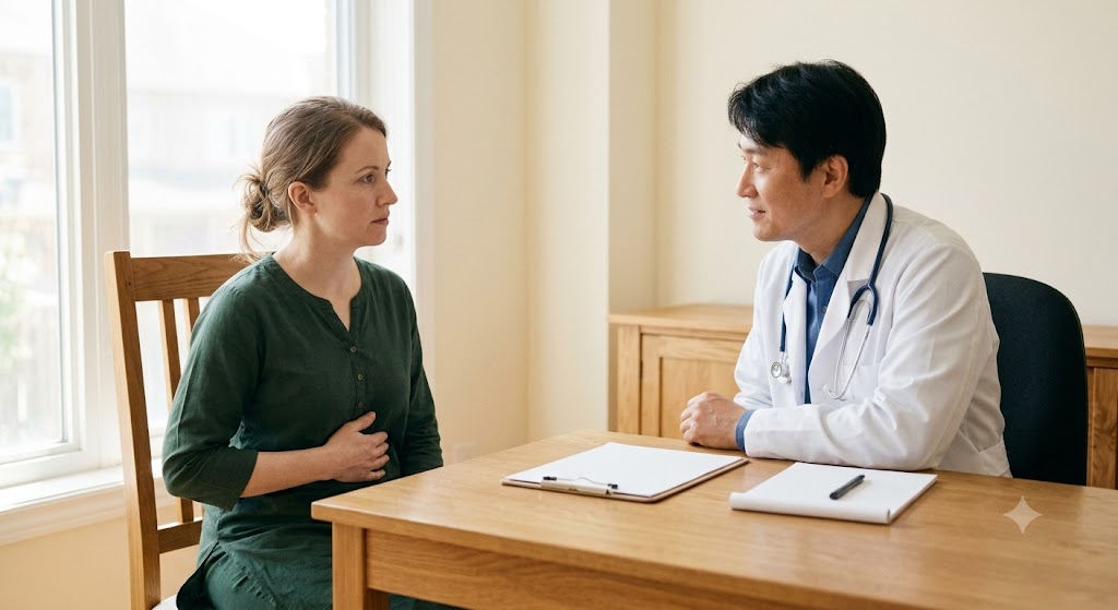
This guide is for adults and athletes who have started to notice stiffness, sounds, catching, or instability in the knee but aren't sure whether it's serious enough to see a doctor. It organizes suspected conditions by symptom to help you gauge for yourself whether a clinic visit is needed, but it is not a tool for confirming a diagnosis. An accurate diagnosis requires a physical examination — where a doctor directly touches and moves the joint to assess its condition — by an orthopedic specialist, and imaging tests if necessary.
Why Knee Pain Happens: Biomechanics and Risk Factors
A Joint Structure That Bears 3–5 Times Body Weight
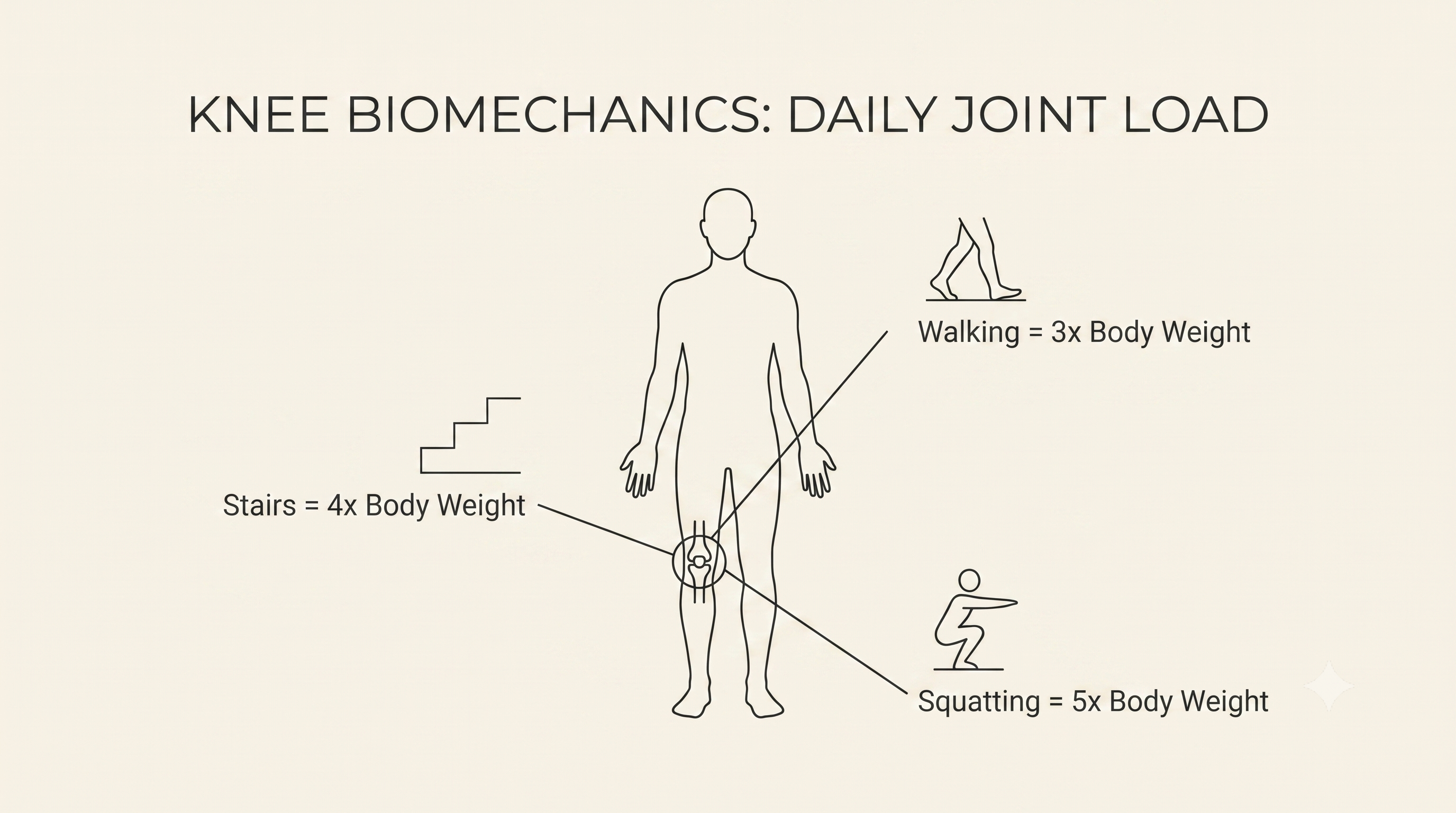
The knee is a weight-bearing joint that repeatedly bears a load of around 3–5 times body weight even during everyday movements like walking and climbing stairs. Walking on flat ground places about 3 times body weight on the knee, while climbing stairs or squatting increases this to as much as 5 times. Whereas the hip joint maintains stability through a ball-and-socket bone structure, the knee instead relies solely on the balance of ligaments, tendons, the meniscus, and cartilage to stay stable, without this kind of bony support. This makes it relatively more vulnerable to trauma and degenerative changes. In addition, cartilage and ligaments receive limited blood flow, so once damaged, their natural capacity to heal is limited — which is why it's important to recognize symptoms early rather than ignore them.
4 Key Risk Factors: Trauma, Aging, Leg Alignment, and Obesity
The risk factors for knee pain generally fall into four categories.
- Trauma and sports injuries: Athletes who repeatedly experience sudden stops, direction changes, and strong impacts
- Age-related degenerative changes: Adults age 50 and older, whose cartilage gradually wears down
- Leg misalignment (bow-leggedness or knock-knees): Cases where load is concentrated on a specific part of the knee
- Obesity: Since the weight-bearing multiplier itself increases, the absolute load on the joint also increases
These four factors can act alone or in combination. For example, in an obese person in their 50s, degenerative changes and increased load act together, compounding the risk. If you're 50 or older, or concerned about degenerative changes, it may help to also review the health screening items recommended by age group, not just for the knee.
👉 Wondering what other health checks make sense for your age group, beyond the knee? See the recommended health screening checklist by age!
6 Knee Conditions Distinguished by Symptoms
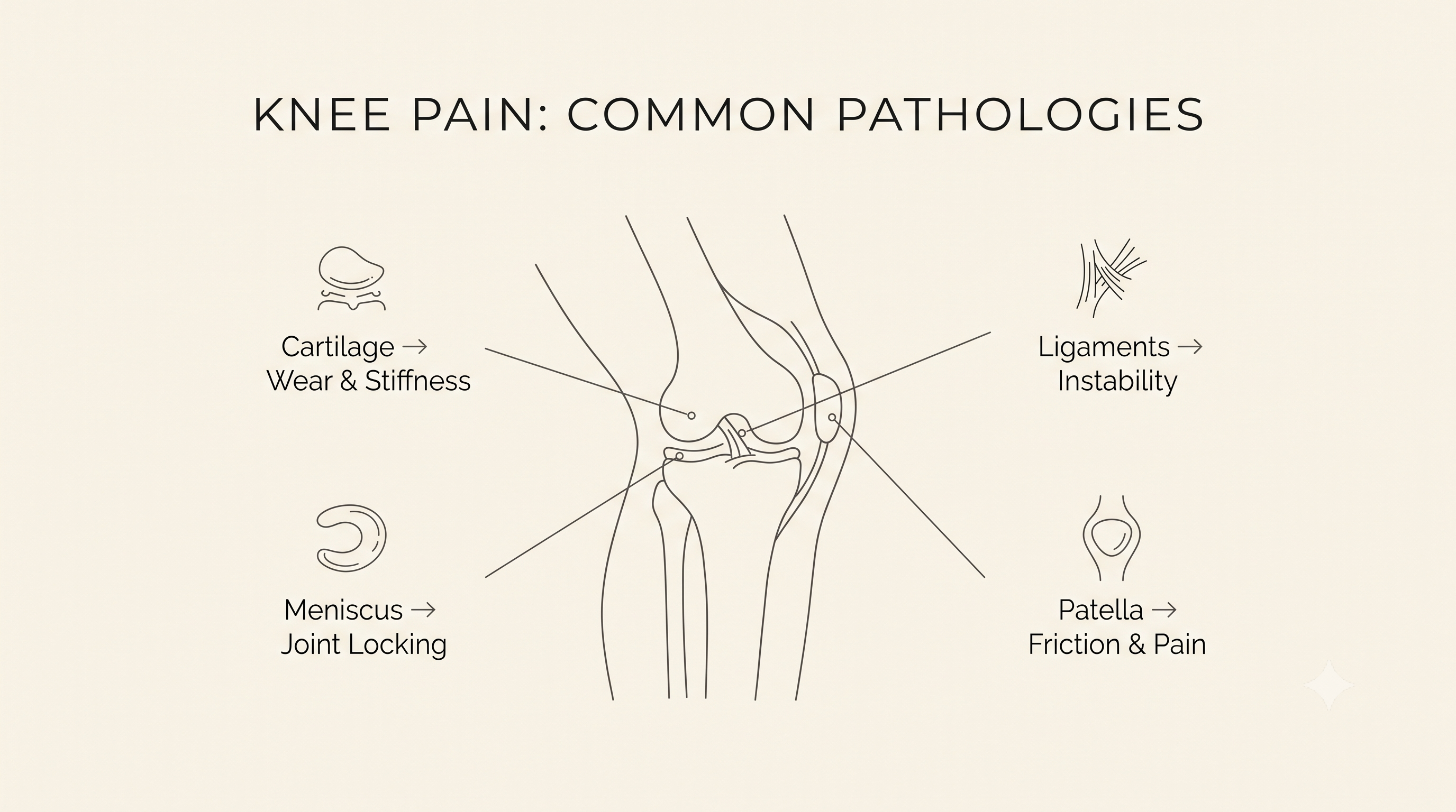
The sounds, catching sensations, and tenderness that appear in the knee follow different patterns depending on the condition. If crepitus (a grinding sound) and morning stiffness recur, degenerative arthritis may be suspected; if the knee catches or doesn't fully straighten, meniscus injury may be suspected. If a "pop" sound is followed by sudden swelling and instability, a cruciate or collateral ligament injury is possible, and localized tenderness and warmth in a specific area are common in tendinitis or bursitis. The "theater sign" — a sharp pain at the front of the knee when going down stairs, similar to standing up after a long movie — is characteristic of chondromalacia patellae, and protrusion and pain at the shinbone in adolescents during growth spurts are likely signs of Osgood-Schlatter disease. However, this classification is for reference only, and it's not uncommon for multiple conditions to occur at once or for symptoms to overlap.
| Condition | Main Cause | High-Risk Group | Key Symptoms |
|---|---|---|---|
| Degenerative arthritis | Gradual wear and degenerative change of joint cartilage | Age 50+, postmenopausal women, obesity | Crepitus (grinding sound), morning stiffness |
| Meniscus injury | Tear or degenerative damage of the meniscus | Direction-change sports, older adults | Joint catching, incomplete extension |
| Knee ligament injury (ACL/PCL/MCL/LCL) | Ligament tear from sudden stops, twisting, or strong impact | High-intensity sports, high-energy trauma | "Pop" sound, sudden swelling, instability |
| Tendinitis/bursitis | Overuse-related inflammation of tendons/bursa | Running, jumping sports, repeated squatting | Localized tenderness, warmth |
| Chondromalacia patellae | Repeated friction and softening of cartilage behind the kneecap | Young adults, runners, muscle imbalance | "Theater sign" (pain going down stairs) |
| Osgood-Schlatter disease | Repeated traction stress on the shinbone growth plate | Adolescents aged 10–15 during growth spurts | Shinbone protrusion, localized pain |
Degenerative Arthritis — Crepitus and Morning Stiffness
As cartilage gradually wears down, bone rubs against bone, producing a grinding sound (crepitus), and stiffness upon waking is a characteristic feature. Risk increases in those age 50 and older, especially in postmenopausal women and those who are obese.
Meniscus Injury — Joint Catching and Buckling
A sudden catching sensation or the feeling that the knee won't fully straighten is typical. It can also appear as a degenerative tear in athletes who frequently change direction or in older adults.
Knee Ligament Injury (ACL, PCL, MCL, LCL) — "Pop" Sound and Instability
A "pop" sound at the moment of injury is often followed by rapid swelling, and a lingering sensation that the knee gives way or buckles frequently remains afterward. This is common in high-intensity sports or high-energy trauma such as car accidents.
Tendinitis/Bursitis — Localized Tenderness and Warmth
Repeated overuse strain on a tendon or bursa causes pain and warmth when a specific area is pressed. Running, jumping exercises, and repeated squatting are risk factors.
Chondromalacia Patellae — The Theater Sign
Repeated friction on the cartilage behind the kneecap makes pain more noticeable when going down stairs or standing up after sitting for a long time. This is common in young adults, runners, and those with a thigh muscle imbalance.
Osgood-Schlatter Disease — Shinbone Protrusion in Adolescents
Repeated traction on the shinbone growth plate causes a bump and pain below the knee. It mainly occurs in adolescents aged 10 to 15 during growth spurts, and in most cases improves once the growth plate closes, though some may retain a protruding bone even into adulthood.

Considerations: The Limits of Self-Diagnosis and Why Differentiation Matters
Some knee conditions are difficult to distinguish by symptoms alone. A notable example is that a torn anterior cruciate ligament (ACL) and a meniscus injury can both cause swelling and restricted movement, making them hard to differentiate through a physical exam alone in some cases — precise imaging tests such as MRI are needed here. In addition, since ligaments and cartilage have limited capacity for natural healing once damaged, leaving them untreated can accelerate joint function decline or degenerative change. If an ACL tear is left untreated, the rate of secondary meniscus injury within 5 years increases substantially to 50–70%, and the rate of early-onset degenerative arthritis within 10–20 years rises to 50–80% or higher; likewise, an untreated meniscus injury raises the risk of degenerative arthritis within 5–10 years to 3–5 times that of the general population. This guide is meant only as a reference to help narrow down suspected conditions based on symptoms — it is not a tool for confirming an actual diagnosis. Diagnosis is made through a physical examination by an orthopedic specialist and, if needed, X-ray or MRI imaging; in particular, for multi-structure injuries where both ligament and meniscus damage are suspected simultaneously, an in-person specialist evaluation should take priority over self-diagnosis.
When Symptoms Alone Aren't Enough to Distinguish (ACL Tear vs. Meniscus Injury)
Both conditions can involve sudden swelling and restricted movement, and there are real cases where palpation or history-taking alone cannot confirm the diagnosis. In these cases, moving on to imaging tests is the safer approach.
The Risk of Leaving It Untreated — Cartilage and Ligaments Have Limited Natural Healing
Cartilage and ligaments are tissues with limited blood supply, so they regenerate slowly even after injury. If you ignore the pain because it seems mild, the damage can accumulate.
Why This Guide Cannot Confirm a Diagnosis
The same symptom can appear across multiple conditions, and because pain thresholds and the degree of damage vary from person to person, using the information in this article alone to make a diagnosis is not recommended.
If You're Seeing a Doctor in Korea: Initial Visit Procedures and Tests
Initial History-Taking and Physical Examination Flow
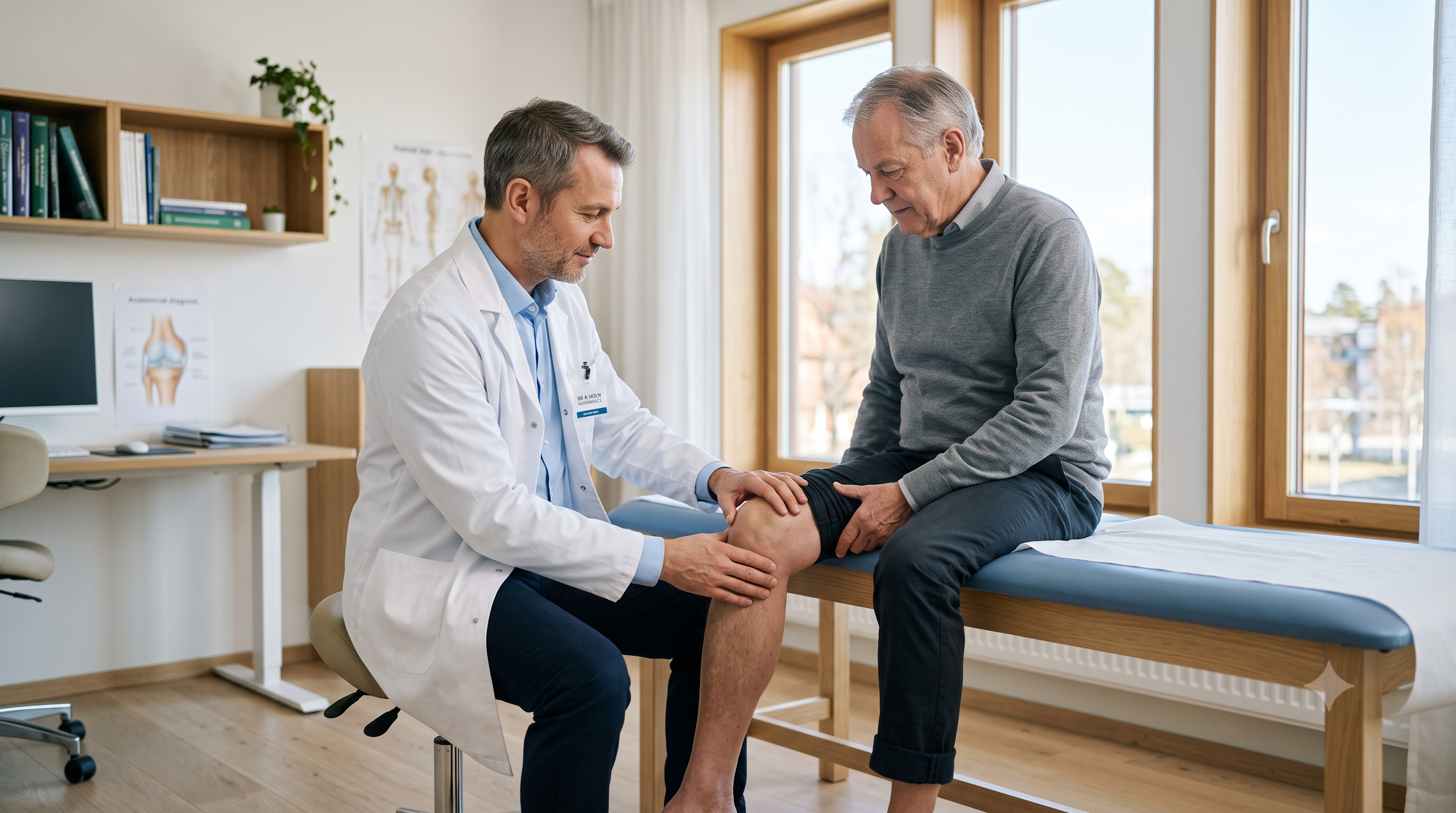
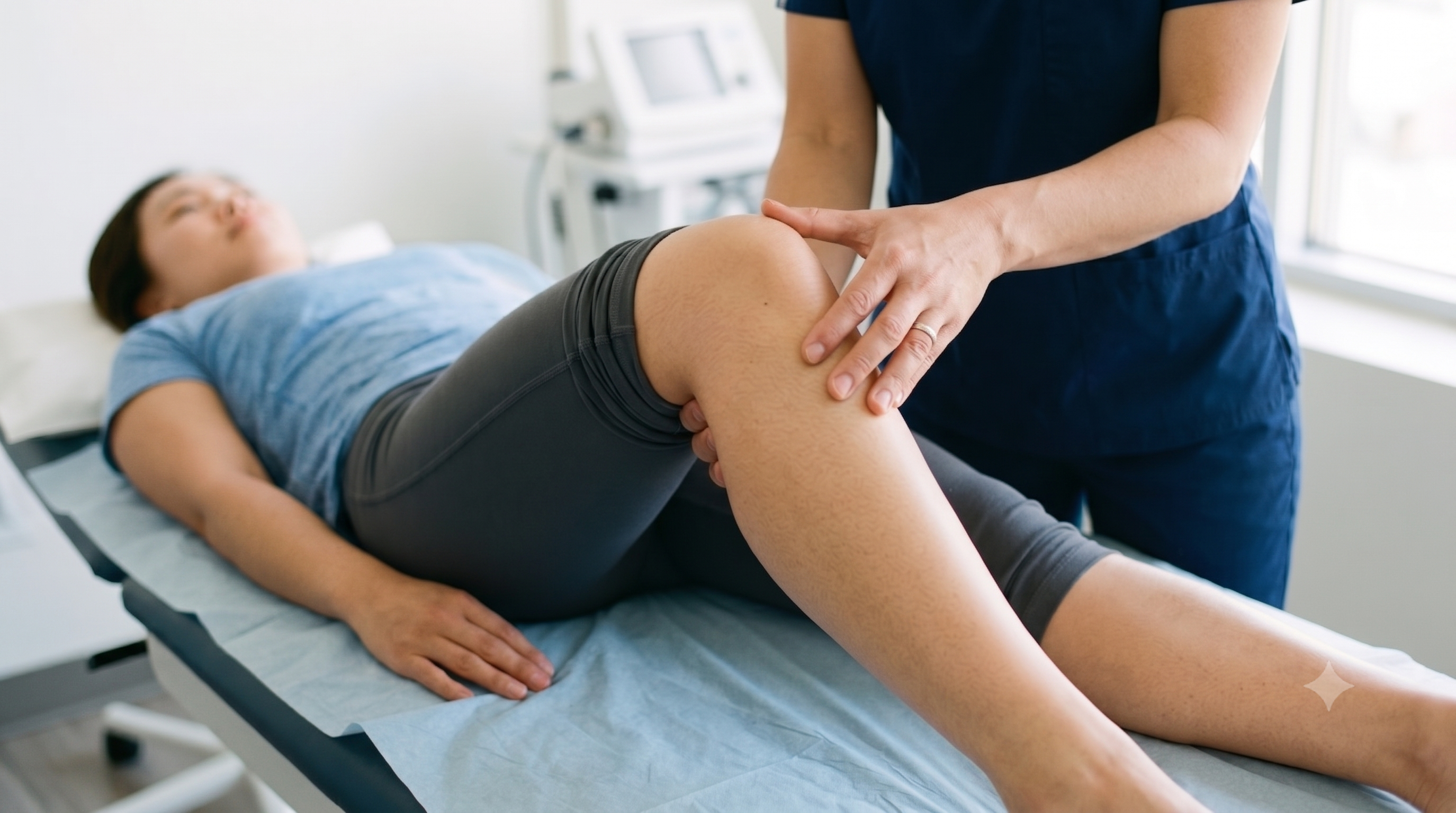
An orthopedic visit typically begins with a history-taking process — asking about the location and onset of pain and any accompanying symptoms — followed by palpation to check range of motion and areas of tenderness, and then special tests to assess ligament stability (such as the Lachman test).
When X-ray or MRI Is Needed
If a bone structural abnormality or a change in joint space is suspected, an X-ray is performed; if soft tissue damage to the ligaments or meniscus is suspected, an MRI is added. The staged treatment options based on test results are covered in detail in a companion guide comparing non-surgical and surgical treatments for knee pain diagnosis. You can also review the general range of tests and treatment options available at orthopedic clinics through a directory of available treatments and procedures.
Number of Initial Visits and Time Required
Typically 1 visit (1–2 visits depending on whether same-day MRI is performed and results reviewed). Time required: approximately 30 minutes to 1 hour for the exam and basic X-ray (approximately 1 hour 30 minutes to 2 hours total if same-day MRI is performed).
Initial Visit Cost Range
These figures are based on non-insured (self-pay) costs for foreign patients without national health insurance coverage; the amount can vary significantly depending on the type of hospital (clinic, general hospital, university hospital) and the applicable fee schedule, so confirming the exact fee with hospital staff is necessary.
Additionally, what to bring and language support vary by hospital when booking an appointment.
- Documents to prepare in advance: Identification (passport or foreign resident registration card), medical records and imaging CDs from other hospitals (if available), an English-language doctor's note or diagnosis certificate (recommended if you have prior treatment history abroad)
- English consultation support: Available (dedicated international care coordinators are provided; confirm availability when booking in advance)
- Recovery/downtime: Not applicable (this part covers the symptom-identification stage; recovery time after treatment is covered separately)
👉 Curious what tests and treatment options are typically available at Korean orthopedic clinics for knee pain? Browse the list of available treatments and procedures!
❓ Frequently Asked Questions (FAQ)
Q. What's the difference between a cruciate ligament tear and a meniscus injury?
A cruciate ligament tear is characterized by a "pop" sound right after the injury followed by rapid swelling and instability, while a meniscus injury commonly causes the knee to catch or fail to straighten.
- How it works: Ligament injury undermines the joint's overall stability, while meniscus injury creates a physical catching sensation during movement
- What to check: Both conditions can be difficult to distinguish by symptoms alone in some cases, so precise imaging such as MRI may be needed
- Cost and time required: Approximately ₩400,000–₩800,000 on a non-insured basis (separate confirmation needed for the foreign-patient fee schedule); approximately 20–40 minutes for imaging of the knee area
Q. My knee makes a "popping" or "crunching" sound — do I definitely need to see a doctor?
Joint sounds without pain or swelling can be a normal physiological phenomenon, but if they're accompanied by pain, swelling, or catching, a medical visit is needed.
- Degenerative arthritis: accompanied by crepitus (grinding sound)
- Chondromalacia patellae: sound and pain occur together when using stairs
- If accompanying symptoms are present, an orthopedic appointment is recommended — general orthopedic clinic: same day to within 2 days; general or university hospital: approximately 1–4 weeks
Q. Why does my knee only hurt when going up or down stairs?
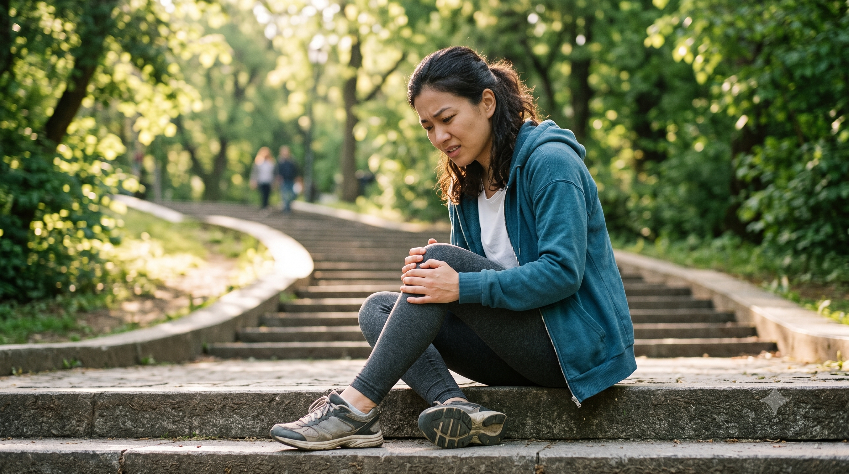
This is because stairs place more load on the knee than walking on flat ground, and pain tends to be more pronounced in those with chondromalacia patellae or degenerative arthritis.
- Chondromalacia patellae: increased pressure on the cartilage behind the kneecap
- Degenerative arthritis: worn cartilage can't sufficiently absorb the load
- What to check: if symptoms persist, it's safer to avoid strenuous exercise and see a specialist
Q. When does knee pain require an emergency room visit?
If you're completely unable to bear weight on the leg, or if you have sudden swelling with fever, or a visible joint deformity, seek emergency care immediately rather than attempting self-care.
- Inability to bear weight
- Acute swelling accompanied by fever, chills, or skin redness (possible joint infection)
- Joint deformity or bone protrusion
- Loss of sensation or persistent numbness in the calf, foot, or ankle
- Recommended visits if these symptoms occur: immediate emergency care, one time
Next Steps — If You Want to Learn About Diagnosis and Treatment
Everything covered here is reference material meant to help narrow down suspected conditions by observing symptoms — the actual diagnosis and treatment decisions must go through a specialist's examination.
Part 2: Go to Knee Pain Diagnosis and Treatment
Whether non-surgical treatment is sufficient or surgical treatment is needed depends on the extent and location of the damage, as determined by test results. The step-by-step breakdown continues in a dedicated guide covering knee pain diagnosis and a comparison of non-surgical versus surgical treatment.
👉 Wondering which treatment path fits your test results — non-surgical care or surgery? Read the guide on knee pain diagnosis and treatment comparison!