Knee Pain Diagnosis and Stepwise Treatment: Non-Surgical vs. Surgical Treatment (Part 2)
💡 Quick Summary: At a Glance
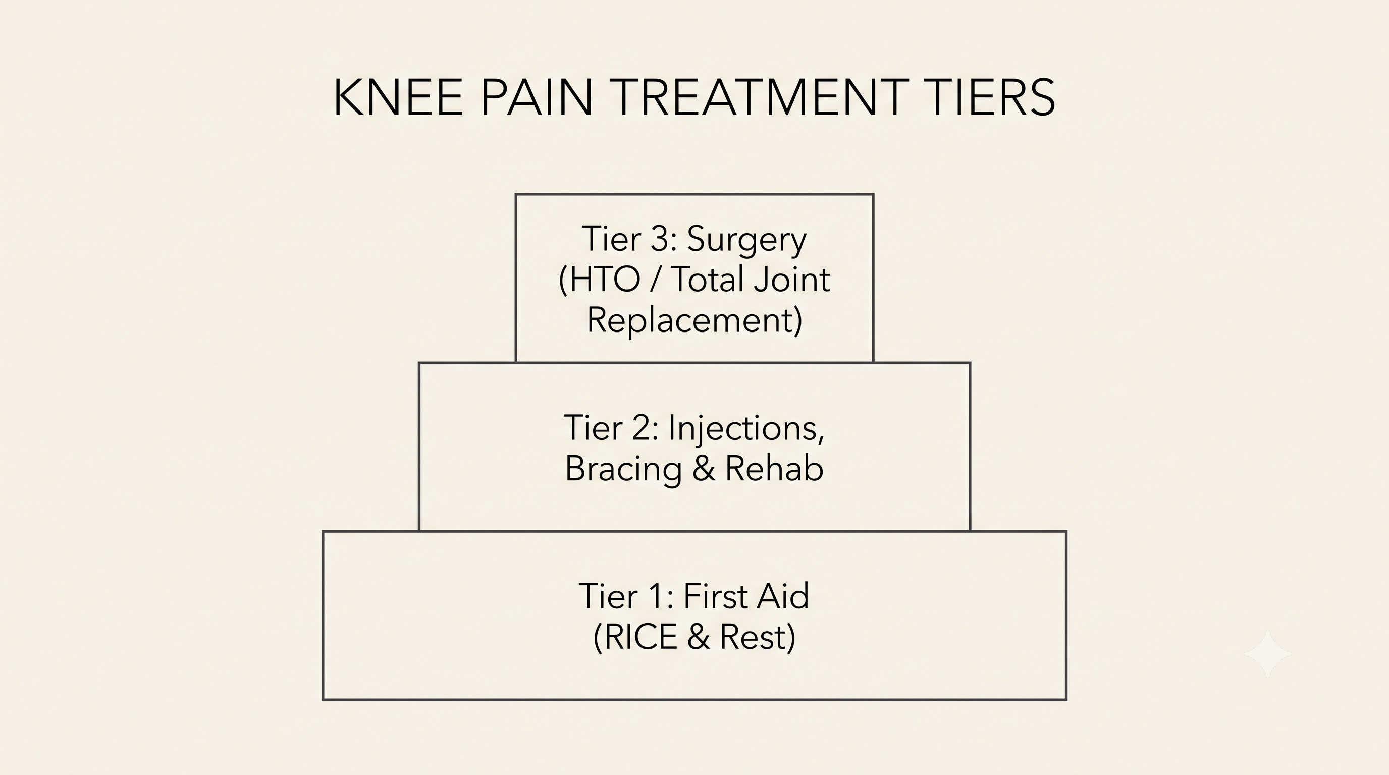
- Stepwise approach: Treatment generally moves from conservative care (rest, ice, activity changes) to non-surgical options (braces, injections, rehab) and, if needed, to surgery (arthroscopy, HTO, TKA), based on the severity of the damage.
- Diagnosis first: X-ray, MRI, and CT are added step by step to confirm the exact cause before deciding which treatment stage is appropriate.
- When to see a specialist right away: Pain lasting 2-3 weeks or more, swelling, instability, catching/locking, or difficulty walking means you should skip self-care and see an orthopedic specialist.
🎯 Who This Guide Is For: Anyone with ongoing knee pain who wants to understand how diagnosis works and how treatment decisions move from conservative care through non-surgical options to surgery.
1. Knee Pain: Accurate Diagnosis Comes First
Diagnosing knee pain begins with taking a medical history and performing a physical examination, followed by imaging tests such as X-ray, MRI, and CT to identify the cause of the pain. Once the cause is identified, treatment generally proceeds in stages: conservative care → non-surgical treatment → surgical treatment. Mild sprains or early pain from overuse often improve with RICE (rest, ice, compression, elevation) and activity modification alone. However, if the damage is moderate or conservative care doesn't bring improvement, non-surgical methods such as braces, anti-inflammatory pain relievers, injections, and rehabilitation are tried next. If pain and functional limitations persist, or the joint damage is severe, surgical treatments such as arthroscopic surgery, high tibial osteotomy (HTO), or total knee arthroplasty (TKA) may be considered.
However, if pain lasts 2-3 weeks or more, or is accompanied by swelling, a feeling of the knee giving way or being unstable, a catching or locking sensation, or difficulty walking, you should skip the step-by-step approach and see an orthopedic specialist right away. These symptoms may indicate damage to a ligament or the meniscus (the crescent-shaped, shock-absorbing cartilage on the inner and outer sides of the knee).
See a Doctor Right Away If You Have These Symptoms
🚨 If you have even one of the symptoms below, see an orthopedic specialist instead of relying on self-care.
- Pain lasts 2-3 weeks or more
- The knee is swollen or feels warm
- There's a sense of instability, such as the knee giving way or feeling weak when walking
- There's a catching or locking sensation
- Walking itself is difficult
If You Haven't Checked the Cause and Symptoms in Part 1 Yet
If you're still not sure what type of pain you're experiencing, start with a guide that organizes information by pain location and the situations that trigger it. This article focuses on what comes after diagnosis — specifically, which tests and treatments follow.
👉 Not sure yet what's causing your knee pain? Check the causes and symptoms of knee pain first
2. How Is Knee Pain Accurately Diagnosed?
X-ray, MRI, and CT each check for different things. X-ray is the basic test that checks bone alignment, whether the joint space has narrowed, and whether there's a fracture or bone spurs (bony growths), and it's typically the first test performed for most knee pain cases. MRI is needed to check soft-tissue damage that X-ray can't show, such as damage to cartilage, the meniscus, ligaments, or tendons, and it's ordered additionally when a ligament tear or meniscus damage is suspected. CT is used mainly at the surgical-planning stage, when a complex fracture or the overall alignment of the leg needs to be analyzed in three dimensions. In other words, not every patient needs all three tests — the typical approach is to add only the tests that are necessary, step by step, based on the medical history and physical examination findings. In rare cases, a bone scan may be performed if a stress fracture or infection is suspected, and diagnostic arthroscopy may be used if the diagnosis remains unclear after imaging alone.
History Taking and Physical Examination
The process begins with taking a history — asking when the pain started and with what movement, whether there was swelling or warmth, and whether there's a history of prior injury — along with a physical exam that involves moving the knee directly to check its stability and range of motion.
The Different Roles of X-ray, MRI, and CT
| Category | What It Checks | When It's Typically Needed |
|---|---|---|
| X-ray | Bone alignment, joint space, fractures, bone spurs | First-line test for most knee pain |
| MRI | Cartilage, meniscus, ligaments, tendons | When soft-tissue damage is suspected |
| CT | Complex fractures, leg alignment, 3D analysis | Surgical planning, evaluating complex fractures |
When a Bone Scan or Diagnostic Arthroscopy Is Needed
A bone scan is added when a stress fracture or infection is suspected but isn't clear on X-ray or MRI. If the cause still remains unclear even after imaging, diagnostic arthroscopy — directly examining inside the joint — may be considered.